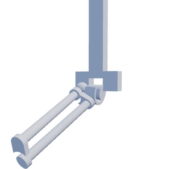
This 3D model is a simplified representation of the skeletal structure of the elbow joint and forearm.
It illustrates how the three joints—the humeroulnar joint, the humeroradial joint, and the proximal radioulnar
joint—contribute to elbow flexion and extension, as well as forearm pronation and supination.
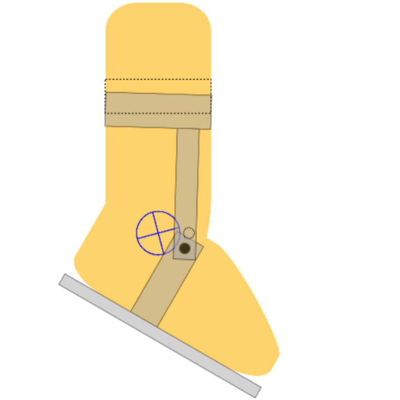
This illustrates how the calf cuff of an ankle-foot orthosis moves during plantarflexion and dorsiflexion
when the orthotic ankle joint is misaligned with the anatomical ankle joint.
i-mobile Banner Ad (Bottom)
About this site
This site introduces tools that use 2D and 3D interactive animations to clearly explain various phenomena.
They are especially well-suited for teachers to use in their classes.
To make these materials easier for teachers to use in class,
detailed explanations have been intentionally left out from the images and models,
and the backgrounds have been kept white so that teachers can add their own notes or explanations.
Privacy & Ads Notice
The advertisements displayed on this site are used to help cover operational costs.
This site uses cookies to improve user convenience and for ad delivery.
Third-party providers may use cookie information to display ads based on users' interests and preferences.